
Eyes are vital organ for one of the five senses, Vision. Let us understand how eye functions to “see”
Let us understand Normal Vision
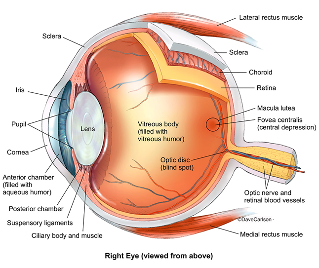
- Light from the objects enters the eye through the cornea, which is the black, dome-shaped extreme front surface of the eye. Cornea is transparent and it has power (which also decides the shape of cornea, like, normal, flat, or steep), the light rays converge at Cornea.
- Behind cornea there is a vascular structure called Iris. The Iris has color depending on pigmentations, like, black, brown, green, blue etc.
- There is hole in centre of Iris which is known as pupil. Pupil controls the amount of the light passing through eye, by constricting or dilating the pupil according to amount of light.
- Behind the Iris, there a Lens, which is also transparent, convex structure and has power. From cornea, through pupil, light rays hit the lens and lens focuses them onto the Retina.
- Eyeball is divided into to two segments. Between Cornea to Lens, it is known as Anterior Segment. And it is filled with clear fluid called Aqueous Humor, which provides nutrition to some part of the eye.
- Natural lens is attached to Ciliary Body by suspensory ligaments. Contraction and relaxation of ciliary body muscle, helps the lens to change the shape to focus near objects. This process is known as Accommodation.
- Posterior segment is a compartment between Lens and Retina, which is filled with transparent jelly like substance called Vitreous Humor.
- From the Lens, the light rays pass through Vitreous Humor and get focused on Retina, forming an inversed image.
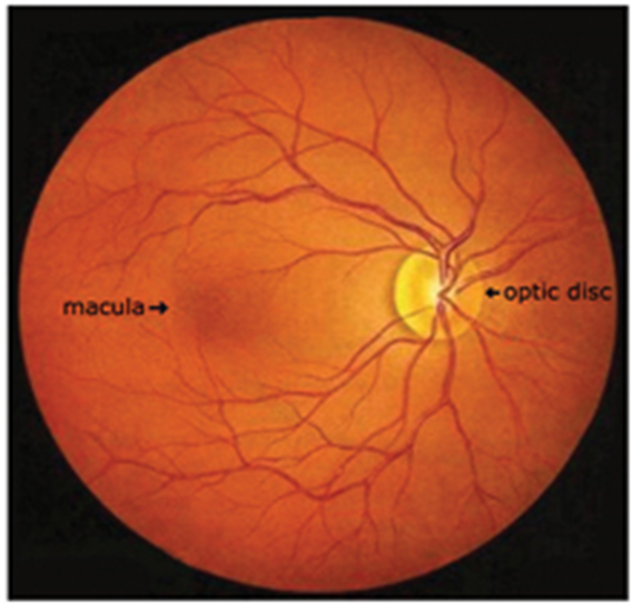
- Retina is innermost, one of the thin three layers on backside of the eye. It is a light sensitive nerve layer. At extreme end, the Retinal nerve fibre layers converge and form a Optic nerve.
- The Optic nerve connects our eyes to brain and is responsible for carrying the visual image signals to the visual cortex of the brain. The visual cortex turns the signals into images and vision is perceived.
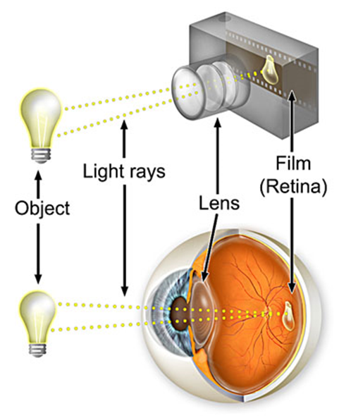
The Normal Eye explained through Camera as an example
The Normal can be very well explained with example of Camera. The eye captures images similar to the way the camera does. The anatomy of the camera bears more similarities to a biological eyeball than many would imagine, including the lens-like cornea and the film-like retina. Similarities like these give the camera the appearance of a robotic eye. However, though there are many similarities between cameras and eyes, they are by no means identical.
Facts About Tears
A happy ending to a story, a sad break-up, an onion chopped into pieces—they all can trigger your tears. Tears serve many purposes, and your eyes produce them all the time. In fact, you make 15 to 30 gallons of tears each year.
Tears are essential to help you see clearly and maintain the health of your eyes. They can also help communicate your emotions.
Your body makes three types of tears.
Basal tears are in your eyes all the time to lubricate, nourish and protect your cornea. Basal tears act as a constant shield between the eye and the rest of the world, keeping dirt and debris away.
Reflex tears are formed when your eyes need to wash away harmful irritants, such as smoke, foreign bodies or onion fumes. Your eyes release them in larger amounts than basal tears, and they may contain more antibodies to help fight bacteria.
Emotional tears are produced in response to joy, sadness, fear, and other emotional states. Some scientists have proposed that emotional tears contain additional hormones and proteins not present in basal or reflex tears.
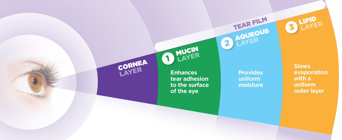
Layers of Tear film:
Tears are not just saline. They have a similar structure to saliva and contain enzymes, lipids, metabolites, and electrolytes.
Each tear has three layers:
- An inner mucus layer that keeps the whole tear fastened to the eye.
- A watery middle layer (the thickest layer) to keep the eye hydrated, repel bacteria and protect the cornea.
- An outer oily layer to keep the surface of the tear smooth for the eye to see through, and to prevent the other layers from evaporating.
Tear Film production:
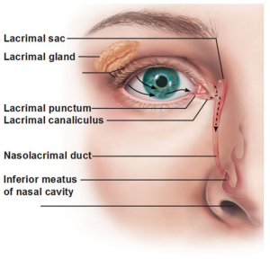
Lacrimal glands above each eye produce your tears. As you blink, tears spread across the surface of the eye. Then the tears drain into puncta, tiny holes in the corners of your upper and lower eyelids. Your tears then travel through small canals in the lids and down a duct before emptying into your nose. There, tears will either evaporate or be reabsorbed.
Sometimes babies are born with a blocked tear duct, a condition that usually resolves on its own.
An eye infection, swelling, injury, age changes over the lid or a tumor can cause a blocked tear duct in adults.
When a lot of emotional or reflex tears are made, they overwhelm the lacrimal drainage system. That is why these tears can spill out of your eyes, run down your cheeks and sometimes dribble out of your nose.
Aging and Tear film:
Basal tear production slows with age, and this can lead to the development of dry eye.
Dry eye is a common problem for people undergoing hormonal changes, especially women during pregnancy and menopause.
Contact lenses and certain medications can also cause dry eye.
Have you noticed, when babies cry, no tear comes out!
If you have dry eye, you may also be prone to blepharitis, a common cause of irritation or swelling of the eyelids.
Our lifestyle, like too much exposure to Digital screens (computers, Laptop, Mobile, IPad/Tablet, Television, Kindle and time on Video games and on social media usage, also causes dry eye, across all age spectrums.